Knee Tendonitis

Knee Tendonitis

Tendonitis of the knee is the inflammation of the tendons that exist around the knee joint, often leading patients to seek out their knee specialist doctor. They are a frequent cause of knee pain and generally result from situations of overstraining, namely those that occur during sports practice, whether in high competition athletes or weekend sportspeople.

The diagnosis is made, primarily, by discriminating the characteristics of the pain, specifying: the location; the inflammatory nature; the triggering factor (usually high intensity exercise) and the response of this condition to anti-inflammatory medication, rest and ice.

Oedema and tumefaction may sometimes be visible in the area affected by tendonitis and palpation of the pain site can help identify the knee structure affected by tendonitis.

Usually, as auxiliary diagnostic tests, ultrasound and MRI of the knee are used, due to their high sensitivity and specificity to identify soft tissue pathology. Radiological examinations may reveal axial deviations of the lower limbs which are also an etiological factor, as well as small calcifications in the tendon insertions (the so-called enthesopathies).

The treatment for knee tendonitis, in general, involves reducing physical activity (or even resting), anti-inflammatory drugs and ice packs.

A rehabilitation programme (physiotherapy) adapted and prepared by your specialist in physical medicine and rehabilitation is important to speed up the process of recovery and eventual return to sport.

Surgery is rarely indicated for the treatment of tendonitis, which is why it should be carefully considered by your orthopaedic surgeon. It is reserved for chronic tendinopathies that are resistant to conservative treatment and have associated partial or complete ruptures.

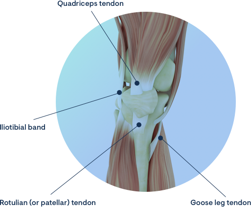
See below specifically which are the most frequent tendonitis/bursitis of the knee and how to treat them individually:

Patellar or Patellar Tendonitis

Rotular tendonitis (or patellar tendonitis) is one of the most common causes of anterior knee pain. It generally occurs in athletes in sports that force them to create an impulse, such as basketball, volleyball and handball, and is therefore referred to as "Jumpers knee". It is associated with high intensity exercise, but also occurs in occasional sportspeople.

The pain caused by patellar or rotular tendonitis can be so intense that it can cause severe functional limitation, leading to the cessation of sporting activity or a reduction in performance level.

In more serious and prolonged cases, chronic inflammation of the patellar tendon may lead to progressive tissue weakening, with the appearance of micro-tears and eventual evolution towards complete and utter rupture of the tendon. In cases which are very resistant to these treatments and which condition great functional incapacity, infiltration with corticosteroids should be carried out with caution in view of the increased risk of tendon rupture, especially when applied multiple times.

Infiltration with PRP's (platelet-derived growth factors) is considered in the literature to have a local analgesic effect and to improve the regeneration process.

The prognosis of Jumpers knee depends on a number of factors such as the time of evolution and intensity of the symptoms, biotype/weight of the athlete, morphology of the knees, type and intensity of the sport practiced. Age also decisively affects recovery time, being obviously shorter in younger athletes.

Surgery is considered a last resort for the treatment of these types of pathologies and consists of debridement of degenerated tissue, cruentation of the lower pole of the patella and, if necessary, reinsertion of any areas of tendon rupture.

Due to the risks of possible complications, namely the complete rupture of the tendon, postoperative recovery should always be very careful, respecting the healing timings of the tissues involved and under the supervision of Orthopaedic and Physiatry.

Quadriceps Tendonitis

Quadriceps tendonitis causes anterior knee pain, more precisely in the region immediately above the patella, the supra-patellar region, where the quadriceps tendon is inserted.

It is caused by excessive exercise and high load, although it more often affects patients at older ages.

Its intensity is variable, but it may evolve to chronicity, with progressive degeneration and eventually rupture, so it must be treated conscientiously with a reduction of activity and a rehabilitation programme appropriate to the athlete and the sport in question.

Iliotibial Band Syndrome

Iliotibial band syndrome is the inflammation of the band located on the lateral aspect of the joint near the insertion area of the tensor fascia lata tendon. It results from friction or rubbing of this structure over the lateral condyle of the femur.

This is one of the most frequent causes of pain or overuse syndrome of the knee in sports medicine (or traumatology) consultations. It particularly affects runners ("runners knee") and cyclists.

Iliotibial band syndrome generally results from performing athletics without adequate stretching of the posterior thigh structures (ischio-tibials) before and at the end of training or competition. Repeated movements with excessive tension on these structures are at the origin of the inflammation. Length differences (dysmetria) or limb misalignment (varus knees), as well as the use of inappropriate footwear, may also contribute to the lateral knee pain which characterises this iliotibial band syndrome. Although it occurs more frequently in athletes, especially in sports involving running, it can also develop in walkers or in patients who do not engage in any regular sports activity.

The main signs and symptoms of this syndrome are pain on the lateral aspect of the knee, which generally appears after a few minutes of running and becomes more intense as the distance covered increases. Some swelling in the affected area may also be visible or palpable.

Your orthopaedic surgeon, a knee specialist, will diagnose iliotibial band syndrome by the type and location of the pain, its onset, usually associated with running, and by a characteristic physical examination, in which pain and crepitus appear on palpation of the iliotibial band near the femoral condyle. The ischiotibial retraction can also be objectified. Magnetic resonance imaging (MRI) of the knee usually confirms the diagnosis by showing a hypersignal area in the affected zone.

In the acute phase, rest, the application of ice and anti-inflammatory drugs are essential for the relief and treatment of the patient's complaints. Physiotherapy acts through the progressive and continuous stretching of the structures of the posterior face of the thigh, cancelling out the retraction of the ischiotibials with specific exercises. The application of local anti-inflammatory agents, or even the use of corticosteroid infiltration, may speed up the recovery process.

Surgery to treat ileotibial band syndrome should be reserved for situations where conservative treatment (physiotherapy) has not been effective. The surgical procedure consists of lengthening the ileotibial band through a Z-shaped plasty of this structure. The correction of lower limb varus can be performed through valgus osteotomy in cases where this predisposing factor is confirmed. It is not a risk or complication-free procedure, so the use of surgery should be properly considered.

Goosefoot Tendonitis or Bursitis

Goosefoot tendonitis is one of the most frequent causes of knee tendonitis in which there is inflammation of the tendons on the medial side of the joint, namely the semitendinosus, semimembranosus and gracilis, which constitute the pes anserinosus. Sometimes the bursa surrounding these tendons is also inflamed giving rise to goosefoot bursitis.

Goosefoot tendonitis is particularly common among women over middle age, with valgus knees, flat feet, some excess weight and who have recently been walking. Other frequent causes of this type of tendinopathy are trauma, osteoarthritis or rheumatic pathology, and even lesions of the adjacent meniscus. Excessive sporting activity ("over training"), such as athletics in climbs, can also lead to its appearance.

The main signs and symptoms are pain in the medial aspect of the knee, especially on going up and down stairs, bending the knee or getting up from a sitting position. Some tumefaction and oedema (swelling) may be visible in the affected area.

Local palpation awakens pain and aids diagnosis. A properly taken clinical history, a careful physical examination, together with auxiliary diagnostic methods such as X-rays of the knee, ultrasound of the knee and Magnetic Resonance Imaging (MRI) of the knee may be essential. They help to identify the causes and visualise the oedema and accumulation of fluid that surrounds the tendon structures and adjacent soft tissues.

In the acute phase, the treatment of goosefoot tendonitis involves: rest; application of ice and taking anti-inflammatory medication (or drugs). Physiotherapy plays a predominant role in the treatment of local inflammation, quadricipital strengthening and sensorimotor training (with an unstable base - BOSU). Corticosteroid infiltration is a fallback treatment, which can provide rapid and often long-lasting relief of symptoms but should be applied in a considered manner by your Orthopaedic or Physiatrist.

Surgery (or operation) is only indicated in cases where the causal factor of the tendonitis can only be corrected by an intervention such as arthroscopy (in cases of meniscal damage) or osteotomy to correct the axis of the knee. Knee arthroplasty is reserved for older patients and those with advanced arthrosis.