Intervention Radiology

Intervention Radiology

Interventional radiology integrates a set of procedures performed without the need for hospitalization (ambulatory), only with local anesthesia, and that usually uses ultrasound control (although it can also be performed with the use of an image intensifier or CT scan) to guide a needle to the site of the lesion by aspirating its contents, injecting drugs or even harvesting a small portion of tissues for biopsy.
 
It is thus possible with minimal discomfort or pain for the patient, aspirating calcifications from the rotator cuff (barbotage of calcifying tendinopathy), treating adhesive capsulites by capsular hydrodistension, infiltrating joints or adjacent tendon structures, treating by dry needling tendinopathies (patella, aquiliana or epicondylianas) or  plantar fasciitis.
 
Modern interventional radiology techniques allow a safe treatment and fast recovery of the patients suffering from the aforementioned pathologies:

Cuff Calcifications

BARBOTAGE / LAVAGE OF SHOULDER ROTATOR CUFF CALCIFICATIONS WITH ULTRASOUND GUIDED NEEDLE

It is a medical technique established in the treatment of calcium tendinopathy, namely calcification of the shoulder rotator cuff, safe, often avoiding surgical procedures and contributing to the earlier recovery of the normal daily activity of the patient.
 
This is an outpatient procedure, with local anesthesia only, which uses ultrasound to guide a fine needle through which saline lavage and aspiration of the calcifications deposited inside the tendons of the rotator hood are performed, thus avoiding exposure to ionizing radiation.
 
With ultrasound control, an experienced operator can direct and real-time visualization of heterotopic calcification, with access to lavage or fragmentation, according to the evolutionary state of calcium tendinopathy, with minimal tendon injury and avoiding the lesion of the tendon structures , articular and ligamentary satellites.
 
Ultrasound allows at the same time, the identification of other associated pathologies, such as bursitis or tendinous ruptures.
 
The technique of barking (of needling) and lavage of calcifications, together with infiltration of the echogenic bursa, improve clinical and imaging results in 1 year in patients with calcium tendinopathy of the rotator cuff.
 
With this technique there is a probability of 60-70% of complete or significant improvement of the symptoms and return to the activity.
 
Sometimes, two treatments may be needed, at intervals of 6 weeks. About 1/3 of patients may not respond to treatment, and may require surgery.

References;
 
Ultrasound-Guided Barbotage in Addition to Ultrasound-Guided Corticosteroid Injection Improved Outcomes in Calcific Tendinitis of the Rotator CuffJ Bone Joint Surg Am, 2014 Feb 19; 96 (4): 335-335. http://dx.doi.org/10.2106/JBJS.9604.ebo887.D.A.T. Silver, P. Dekimpe and T.D. Bunker.
 
Calcific Tendonitis Of The Shoulder; Is There A Place For Ultrasound Guided Barbotage Or Extracorporeal Shockwave Therapy As An Alternative To Surgery? Journal of Bone and Joint Surgery - British Volume, Orthopedic Proceedings. Vol 84-B, Issue SUPP II, 193.


Echodistension of the articular capsule in adhesive capsulitis of the shoulder

ECHODISTENSION OF THE ARTICULAR CAPSULE IN ADHESIVE CAPSULITIS OF THE SHOULDER

The adhesive capsulitis of the shoulder is a clinical entity that is characterized by pain and stiffness of this joint. The pain, usually intense, results from an inflammatory condition that reaches the joint capsule, thickening it and causing retraction and adhesions, with progressive stiffness.
 
It is usually treated conservatively with medication and an adequate rehabilitation program that can be extended in time for a few months.
 
The echogenic hydrodistension of the joint capsule is a procedure sometimes necessary to control pain and accelerate the recovery of shoulder mobility. It involves the distension of the capsule by the injection of a mixture of anesthetic, serum and corticosteroid, allowing the release of the capsular adhesions created by the inflammation.
 
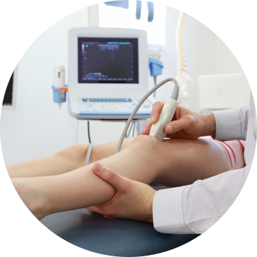
It is a simple and safe procedure, usually taking no more than 10 minutes, performed in an outpatient clinic, ideally by an echo-guided technique, to avoid unnecessary use of ionizing radiation. It allows the visualization of the procedure in real time, with confirmation of the correct positioning of the needle, with greater success rates, and the reduction of the risk of injury of the neighboring structures.
 
There is evidence that capsular distension allows relief of pain, improvement of range of motion and functionality in cases of adhesive capsulitis that does not respond to medication and physiotherapy instituted.
 
Studies also suggest that distension with serum + steroids seems to be more efficient than an isolated corticosteroid injection. It may even be as effective as manipulation over general anesthesia, but with less risk and complication rate.
 
It has a success rate of more than 70% in the reacquisition of range of motion, as well as a rate of over 90% in pain improvement when the procedure is performed by an experienced specialist physician.
 
References:

The role of capsular distention in adhesive capsulitis. Archives of physical medicine. 2003; Gavant ML, Rizk TE, Gold RE, Flick PA. Jacobs LG, , Smith MG, Khan SA, Smith K.

Manipulation or intra-articular steroids in the management of adhesive capsulitis of the shoulder? A prospective randomized trial. Journal of shoulder and Elbow 2009; Quraishi NA, Johnston P, Bayer.

Thawing the frozen shoulder to randomized trial comparing manipulation under anesthesia with hydrodilatation. Journal of Bone and Surgery 2007 Ng CY, Min AK, McMullan L, McKie, S, Brenkel IJ, Cook RE. A prospective randomized trial comparing manipulation under anesthesia and capsular distension for the treatment of adhesive capsulitis of the shoulder. Shoulder and Elbow. 4 (2) 95-99. 2012).
 

Echoed, peritendinous and articular bursae joint infiltration in acute inflammatory conditions

ECHOED, PERITENDINOUS AND ARTICULAR BURSAE JOINT INFILTRATION IN ACUTE INFLAMATORY CONDITIONS
 
The echo-guided joint injection allows a greater precision and control of the exact location in which we intend to infiltrate the drug. It also makes a previous evaluation of the articular pathology, with the exclusion of potential associated complications, and also allowing comparison with the contralateral side.
 
Ultrasound documents the positioning of the needle in real time, drastically reducing the possibility of injury to surrounding structures, such as tendons and ligaments, as well as the risk of vascular lesions, such as bruising or possible pseudo arterial aneurysm formations.
 
If the steroid is not injected correctly into the joint or tendinous sheath, there is a possibility of tendon rupture and cytoske- toato-necrosis of adipose tissues.
 
This technique also allows aspiration of intra-articular contents, lumen of bursae, or tendinous sheaths, with delivery of the material for analysis, namely microbiology.
 
The technique to be performed depends on the experience of the physician, the joint in question and the underlying or associated pathology. Here, too, there are benefits of ultrasound associated with puncture, since it allows adaptation to the specific joint and its particularities, namely joint deformities present in some chronic patients.

Ultrasound-guided synovial biopsy

ULTRASOUND-GUIDED SYNOVIAL BIOPSY
 
The performance of echocardiographic synovial biopsy is a minimally invasive technique for synovial tissue collection, being safe and well tolerated by patients with rare complications.
 
It avoids the costly costs of the biopsy performed in the operative block, reducing the patient's morbidity, being a technique that can be performed in an outpatient clinic.
 
Synovial biopsies can allow histological examinations of RNA among others, identifying tissue lesions and monitoring early stages of rheumatoid arthritis or other joint pathologies.

Infiltration of neuromas from Morton

INFILTRATION OF NEUROMAS FROM MORTON
 
Morton's neuromas are the most frequent orthopedic pathologies at the foot level. They are a cause of pain in the anterior plantar region (metatarsalgias) and result of thickening of the interdigital nerves by repeated traumatisms and areas of hyperpressure. They are often associated with halux valgus (commonly referred to as bunion). Discharge of the metatarsal head with special orthoses or insoles and infiltrations often prevent the need for surgery for the excision of Morton's neuroma.
 
The echogenic infiltration technique of Morton's nevromas allows the confirmation of the correct site to infiltrate, which is reflected in the improvement of clinical outcomes. 
 
It allows to evaluate and to identify in real time the existence of other comorbidities, with possibility of immediate treatment. It also prevents accidental infiltration of neighboring structures and injury of adjacent ligament / tendon structures.
 
Due to the clinical success, often achieved by these minimally invasive techniques, performed in an outpatient clinic, some surgeries are sometimes avoided, with the costs and risks inherent to the surgical procedure.
 
References:

Bennett GL, Graham CE, Mauldin DM. Morton's interdigital neuroma: a comprehensive treatment protocol. Foot Ankle Int. 1995; 16: 760-763 .; Hughes RJ, Ali K, Jones H, et al.

Treatment of Morton's neuroma with alcohol injection under sonographic guidance: follow-up of 101 cases. Am J Roentgenol. 2007; 188: 1535-1539. Saygi B, Yildirim Y, Saygi EK, et al.

Morton's neuroma: comparative results of two conservative methods. Foot Ankle Int. 2005; 26: 556-559.
 

Dry needling of tendinopathy

DRY NEEDLING OF TENDINOPATHY:EPICONDYLITIS, PATELLAR, AQUILIAN OR OTHER TENDINOPHATIES
 
Dry needling is a tendon needle piercing technique with the aim of decompressing, stretching the tendon by multiple partial tenotomies, and provoking local stimulation of regeneration and improvement of the local circulation.
 
Echo-guided tenotomy (dry needling with ultrasound control) allows direct visualization of target tissues, improving efficacy and preventing injury to adjacent structures.
 
Studies in the treatment of certain tendinopathies, such as those of Mcshane et al, describe success rates close to those of surgery, with less inherent risks, less invasiveness, and requiring only a local anesthetic.
 
Dry needling stimulates a healing response via local inflammation, leading to recruitment of blood vessels and collagen precursors. This response, when aided by an adequate physiotherapy protocol, induces tendon remodeling.
 
There is still no consensus about the partial tenotomies performed with corticoid injection. In the literature, there are studies that report the following results: co-administration of corticosteroids - excellent results up to 12 weeks, but long-term pain relapses; without corticoid - takes longer to relieve pain, but long-term relief has proven to be excellent.
 
This technique also allows the possibility of injection of sclerosing substances such as 20% dextrose, autologous blood injection or even platelet growth factors (PRPs), with great precision, particularly in small interstitial breaks, which increases its rate of classical infiltration methods.
 

Dry needling of plantar fasciitis

DRY NEEDLING OF PLANTAR FASCIITIS
 
Plantar fasciitis results from inflammation and thickening of the plantar fascia of the foot and that has its insertion in the calcaneus. Calcification of this region can sometimes be seen in the RX, giving rise to what is called the spur of the calcaneum. It affects about 10% of the population and is a painful and disabling condition that can be difficult to treat in adults.
 
Dry needling is an ambulatory, echo-guided, safe, tenotomy technique for the induction of bleeding and the release of scar tissue. 
 
For this reason this technique, because it is fast, minimally invasive, with low costs) may have its indications.
 
In a preliminary study (developed by the University of Genova, Italy), which included 44 patients, a success rate of 95% was achieved in the resolution of symptoms involving plantar fasciitis in 2-3 weeks. Relief of the disease lasted for an average of more than 8 months after the procedure. It is, therefore, a technique to be considered, especially in cases of reluctance to conservative treatment.