Knee Anatomy
THE KNEE

LIGAMENTS


The menisci are fibrocartilaginous structures that distribute the body weight across the knee. They help stabilise the knee as they absorb shock and provide nutrition to the articular cartilage by spreading a thin layer of synovial fluid throughout the joint. There are two menisci in each knee, the medial and the lateral meniscus..
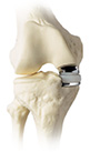
The knee is a complex joint formed by the femorotibial joint (distal femur and proximal tibia) and the patellofemoral joint (femur and patella).
It is also formed by ligaments that stabilise the joint, aided by the menisci that help absorb shock in the cartilages.
The key to a healthy knee is stability and a good alignment of the limb.
LIGAMENTS
The ligaments play a major role in the stability of the posterior and anterior translation, varus-valgus angulation and rotational movements of the knee joint.
The anterior cruciate ligament (ACL) is the main stabiliser of the anterior tibial translation, being the key structure in the daily life of a healthy and active person in sports.
MENISCI
The menisci are fibrocartilaginous structures that distribute the body weight across the knee. They help stabilise the knee as they absorb shock and provide nutrition to the articular cartilage by spreading a thin layer of synovial fluid throughout the joint. There are two menisci in each knee, the medial and the lateral meniscus..


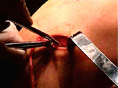

.jpg) Posterior cruciate ligament tears are rare. These injuries are often complex, since they might be associated with injuries in other structures, such as the capsule and other posterior or external structures.
Posterior cruciate ligament tears are rare. These injuries are often complex, since they might be associated with injuries in other structures, such as the capsule and other posterior or external structures.
.jpg)

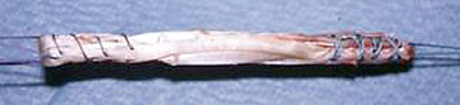
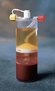 Recently, there have been attempts to use platelet-derived growth factor (PDGF) and chondrocytes culture (MACI) to treat these pathologies, techniques that we have been testing along with the use of stem cells.
.jpg)
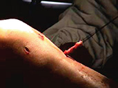
.jpg) The symptoms and the results of the exams will help determine the course of treatment.
The symptoms and the results of the exams will help determine the course of treatment.
.jpg)